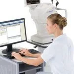
At Hi Def Eyecare Center we utilize the latest Electronic Medical Record (EMR) Software and retinal fundus camera to provide you the best medical and vision care. As part of your comprehensive eye exam we recommend taking a retinal photo. This photo provides information about the general health of your body in addition to your ocular health. It is an invaluable tool to monitor for changes in diabetes and high blood pressure over time as well as eye diseases like glaucoma and macular degeneration.
At over $200k invested in advanced technology, we may be the only Optometry practice in Southern Maryland that has all this Advanced Testing Equipment.
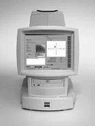
Digital Retinal Imaging & OCT Scans
We use cutting-edge digital imaging technology to assess your eyes. Many eye diseases, if detected at an early stage, can be treated successfully without total loss of vision. Your retinal Images will be stored electronically. This gives the eye doctor a permanent record of the condition and state of your retina.
This is very important in assisting your Optometrist to detect and measure any changes to your retina each time you get your eyes examined, as many eye conditions, such as glaucoma, diabetic retinopathy and macular degeneration are diagnosed by detecting changes over time.
The advantages of digital imaging include:
- Quick, safe, non-invasive and painless
- Provides detailed images of your retina and sub-surface of your eyes
- Provides instant, direct imaging of the form and structure of eye tissue
- Image resolution is extremely high quality
- Uses eye-safe near-infra-red light
- No patient prep required
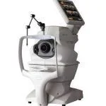
Digital Retinal Imaging
Digital Retinal Imaging allows your eye doctor to evaluate the health of the back of your eye, the retina. It is critical to confirm the health of the retina, optic nerve and other retinal structures. The digital camera snaps a high-resolution digital picture of your retina. This picture clearly shows the health of your eyes and is used as a baseline to track any changes in your eyes in future eye examinations.
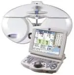
Optical Coherence Tomography (OCT)
An Optical Coherence Tomography scan (commonly referred to as an OCT scan) is the latest advancement in imaging technology. Similar to ultrasound, this diagnostic technique employs light rather than sound waves to achieve higher resolution pictures of the structural layers of the back of the eye.
A scanning laser used to analyze the layers of the retina and optic nerve for any signs of eye disease, similar to an CT scan of the eye. It works using light without radiation, and is essential for early diagnosis of glaucoma, macular degeneration and diabetic retinal disease.
With an OCT scan, doctors are provided with color-coded, cross-sectional images of the retina. These detailed images are revolutionizing early detection and treatment of eye conditions such as wet and dry age-related macular degeneration, glaucoma, retinal detachment and diabetic retinopathy.
An OCT scan is a noninvasive, painless test. It is performed in about 10 minutes right in our office. Feel free to contact our office to inquire about an OCT at your next appointment.
Visual Field Testing
A visual field test measures the range of your peripheral or “side” vision to assess whether you have any blind spots (scotomas), peripheral vision loss or visual field abnormalities. It is a straightforward and painless test that does not involve eye drops but does involve the patient's ability to understand and follow instructions.

An initial visual field screening can be carried out by the optometrist by asking you to keep your gaze fixed on a central object, covering one eye and having you describe what you see at the periphery of your field of view. For a more comprehensive assessment, special equipment might be used to test your visual field. In one such test, you place your chin on a chin rest and look ahead. Lights are flashed on, and you have to press a button whenever you see the light. The lights are bright or dim at different stages of the test. Some of the flashes are purely to check you are concentrating. Each eye is tested separately and the entire test takes 15-45 minutes. These machines can create a computerized map out your visual field to identify if and where you have any deficiencies.
Marco TRS 5100 Total Refraction System
The TRS 5100 benefits from Marco’s latest generation of electronic refraction technology. Replacing the standard refractor, it allows us to control the entire refraction process from a keypad. This keypad also controls the Marco Automatic Chart Projector. And because the TRS 5100 is completely programmable, all the lenses are moved at the touch of a button, taking us to each new refraction step in more efficient and accurate way. The new technology allows you to spend less time in the exam room, and allows us to get a more accurate refraction. The Marco LCD Acuity chart also allow us to have better results for children who can not read letters yet.